Human Bone Anatomy - Teamlabbody 3d Motion Human Anatomy. The humerus or one of the other bones in the shoulder slips out of position. The body's shape is determined by a strong skeleton made of bone and cartilage, surrounded by fat, muscle, connective tissue, organs, and other structures. 2021 newest human model of skeleton for anatomy 67 high with 200+ bones structures,scientific disarticulated human model of skeleton bundle for anatomy, full size male skeleton models with poster,skull, bones, articulated hand & foot. The skeleton acts as a scaffold by providing support and protection for the soft tissues that make up the rest of the body. Bones of the pelvis and lower back the bones of the pelvis and lower back work together to support the body's weight, anchor the abdominal and hip muscles, and protect the delicate vital organs of the vertebral and abdominopelvic cavities.
Produces a collection of high quality casts of the human skeleton models for educators and practitioners in medicine, physical therapy, physical anthropology, comparative anatomy, and biomechanics. Skull, skeletal framework of the head of vertebrates, composed of bones or cartilage, which form a unit that protects the brain and some sense organs. Learn more about the anatomy and function of the skull in humans and other vertebrates. A forearm bone, it runs from the elbow to the thumb. The human skeleton consists of 213 bones, of which 126 are part of the appendicular skeleton, 74 are part of the axial skeleton, and 6 are part of the auditory ossicles.
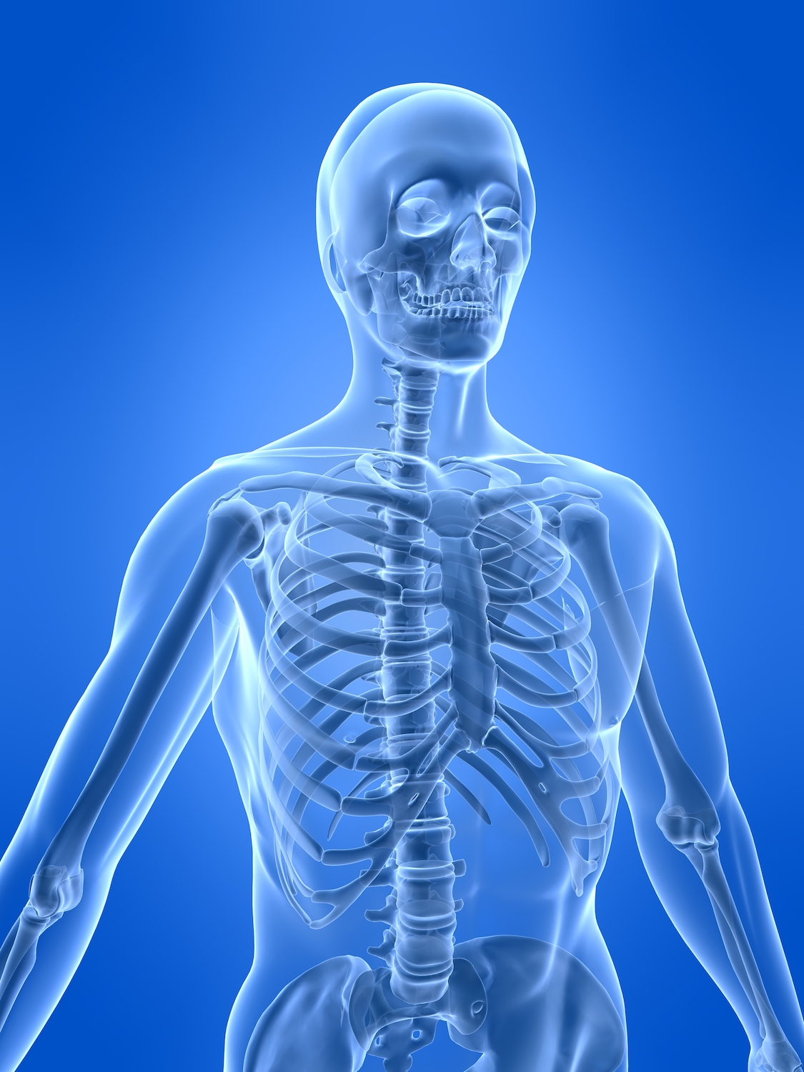
Skeletal System Definition Function And Parts Biology Dictionary from biologydictionary.net In your anatomy & physiology lecture and lab class, you will be required to name each individual bone in the human body. The vertebral column of the lower back includes the five lumbar vertebrae, the sacrum, and the coccyx. The humerus or one of the other bones in the shoulder slips out of position. Quiz on human bones for anatomy & physiology this quiz on human bones is designed to test your knowledge on the location of each individual bone. Learn human bones for anatomy class by using these easy memory tricks (mnemonics)!quiz on human bones: For anatomy students and medical students, it's important to note that this skeleton's right forearm is rotated forward to show how the arm bones look from a different angle. The body's shape is determined by a strong skeleton made of bone and cartilage, surrounded by fat, muscle, connective tissue, organs, and other structures. The skeletal system also provides attachment points for muscles to allow movements at the joints.
In your anatomy & physiology lecture and lab class, you will be required to name each individual bone in the human body.
1 bone is a solid organ that appears pinkish white externally and deep red internally when in a fresh state. Bones of the pelvis and lower back the bones of the pelvis and lower back work together to support the body's weight, anchor the abdominal and hip muscles, and protect the delicate vital organs of the vertebral and abdominopelvic cavities. Learn more about the anatomy and function of the skull in humans and other vertebrates. The knee joins the thigh bone (femur) to the shin bone (tibia). ★ you can rotate models to any angle. Human anatomy is the study of the shape and form of the human body. Quiz on human bones for anatomy & physiology this quiz on human bones is designed to test your knowledge on the location of each individual bone. The smaller bone that runs alongside the tibia (fibula) and the kneecap (patella) are the other bones that make the knee joint. Altogether, the skeleton makes up about 20 percent of a person's body weight. There also are bands of fibrous connective tissue —the ligaments and the tendons —in intimate relationship with the parts of the skeleton. It provides structure to the body, and each bone has a distinct purpose. This is a table of muscles of the human anatomy. The humerus or one of the other bones in the shoulder slips out of position.
Skull, skeletal framework of the head of vertebrates, composed of bones or cartilage, which form a unit that protects the brain and some sense organs. The vertebral column of the lower back includes the five lumbar vertebrae, the sacrum, and the coccyx. Muscles and ligaments pull on the bones of the skeleton at joints to make the body move. This science quiz game will help you learn 15 of the most important bones. The large bones of the arm include:
The Human Skeletal System Live Science from cdn.mos.cms.futurecdn.net A forearm bone, it runs from the elbow to the thumb. The skeletal system contains the bones that give structure to the human body. The human skeletal system consists of all of the bones, cartilage, tendons, and ligaments in the body. The knee joins the thigh bone (femur) to the shin bone (tibia). Learn more about the anatomy and function of the skull in humans and other vertebrates. This diagram depicts skeletal images 744×1314 with parts and labels. This framework consists of many individual bones and cartilages. ★ you can rotate models to any angle.
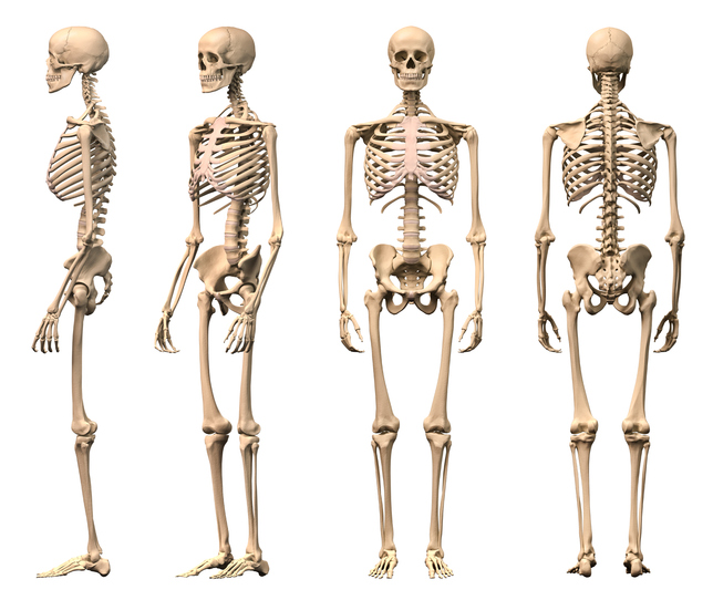
The skeleton of an adult human is made up of 206 bones of many different shapes and sizes.
The skeleton acts as a scaffold by providing support and protection for the soft tissues that make up the rest of the body. Long bone, short bone, flat bone, irregular bone and sesmoid bones. The skull includes the upper jaw and the cranium. The skeleton of an adult human is made up of 206 bones of many different shapes and sizes. The human skeleton of an adult consists of around 206 to 213 bones, and there are 300 bones in children, depending on the counting of sternum (which may alternatively be included as the manubrium, body of sternum, and the xiphoid process). He talks about the anatomy of the. Altogether, the skeleton makes up about 20 percent of a person's body weight. This is a table of muscles of the human anatomy. Reviewed by carol dersarkissian, md on may 18, 2019. Quiz on human bones for anatomy & physiology this quiz on human bones is designed to test your knowledge on the location of each individual bone. Human anatomy bone clones, inc. 4.8 out of 5 stars 29. Bones of the pelvis and lower back the bones of the pelvis and lower back work together to support the body's weight, anchor the abdominal and hip muscles, and protect the delicate vital organs of the vertebral and abdominopelvic cavities.
Reviewed by carol dersarkissian, md on may 18, 2019. The body's shape is determined by a strong skeleton made of bone and cartilage, surrounded by fat, muscle, connective tissue, organs, and other structures. 2021 newest human model of skeleton for anatomy 67 high with 200+ bones structures,scientific disarticulated human model of skeleton bundle for anatomy, full size male skeleton models with poster,skull, bones, articulated hand & foot. The diaphysis and the epiphysis.the diaphysis is the tubular shaft that runs between the proximal and distal ends of the bone. Each bone is a complex living organ that is made up of many cells, protein fibers, and minerals.
No Bones About It The Human Skeleton Is Fascinating Portable Press from www.portablepress.com Bones of the pelvis and lower back the bones of the pelvis and lower back work together to support the body's weight, anchor the abdominal and hip muscles, and protect the delicate vital organs of the vertebral and abdominopelvic cavities. The skeleton of an adult human is made up of 206 bones of many different shapes and sizes. 3d bones and organs (anatomy) a true and totally 3d free app for learning human anatomy with position quiz, built on an advanced interactive 3d touch interface. ★ you can rotate models to any angle. He talks about the anatomy of the. It is composed of 300 bones at birth, but later decreases to 80 bones in the axial skeleton and 126 bones in the appendicular skeleton. The smaller bone that runs alongside the tibia (fibula) and the kneecap (patella) are the other bones that make the knee joint. Bones of the human skeletal system are categorized by their shape and function into five types.
The frontal bone is a flat bone.
Altogether, the skeleton makes up about 20 percent of a person's body weight. Some, like the rib cage, provide protection for softer body parts, while other bones enable mobility by supporting the muscles. It has every bone and organ in the human body. Bones of the human skeletal system are categorized by their shape and function into five types. It provides structure to the body, and each bone has a distinct purpose. Produces a collection of high quality casts of the human skeleton models for educators and practitioners in medicine, physical therapy, physical anthropology, comparative anatomy, and biomechanics. In your anatomy & physiology lecture and lab class, you will be required to name each individual bone in the human body. The body's shape is determined by a strong skeleton made of bone and cartilage, surrounded by fat, muscle, connective tissue, organs, and other structures. The human skeleton consists of 213 bones, of which 126 are part of the appendicular skeleton, 74 are part of the axial skeleton, and 6 are part of the auditory ossicles. Learn more about the anatomy and function of the skull in humans and other vertebrates. The diaphysis and the epiphysis.the diaphysis is the tubular shaft that runs between the proximal and distal ends of the bone. 4.8 out of 5 stars 29. It is composed of 300 bones at birth, but later decreases to 80 bones in the axial skeleton and 126 bones in the appendicular skeleton.